Article on the topic
Enlighten me! Who needs a mammogram?
Such an attitude towards the most delicate area of the female body is unacceptable. After all, even painful tension in the chest before menstruation, small lumps and slight discharge from the nipples are a reason to consult a mammologist. By the way, problems with the thyroid gland, adrenal glands, stress, taking hormonal medications and contraceptives can also affect the condition of the bust. In a word, in order not to face such a serious diagnosis as breast cancer, start preventing this dangerous disease right now.
Pay attention to your diet
Healthy breasts require giving up not only cigarettes, but also an extra cup of coffee and alcohol. The most you can afford is a glass of red wine with lunch. The rest is unnecessary risk. Include foods rich in vitamin C in your diet - lemons, kiwi, bell peppers, cabbage. You should not give up carrots, tomatoes, dill and spinach, which contain vitamin A - it increases antitumor resistance. Vegetable oil and cereals, thanks to vitamin E, prevent or inhibit the appearance of tumors and slow down the aging process. The trace element selenium plays an important role in the prevention of breast cancer. It is able to selectively accumulate in tumor cells and prevent their destructive activity. Therefore, include seeds, garlic, shrimp and fish in your diet.
Skin symptoms in breast cancer and their significance for diagnosis
02.09.2013
Breast diseases
Oncology
As the extensive experience of many researchers shows, skin symptoms are very valuable in the early diagnosis of breast cancer. Sometimes unclear palpation data in combination with skin symptoms make it possible to make the correct diagnosis of breast cancer.
The diagnosis of early forms of breast cancer is made more likely on the basis of skin symptoms, i.e., on the basis of varying degrees of impaired skin mobility in the area of the tumor. It should be noted that the diagnostic value of skin symptoms lies in the fact that they are inherent in the early stages of breast cancer.
During the period of its dynamic development, breast cancer causes varying degrees of impaired skin mobility. It’s like a whole range of symptoms associated with the same cause, but manifesting themselves at different periods of the existence of cancer with different strengths.
It must be assumed that this is due not only to purely mechanical factors, but also to biochemical changes in the tissues surrounding the tumor and depending on the characteristics of tumor metabolism.
The mechanical aspects that cause impaired skin mobility in the tumor area in breast cancer can be easily imagined in the light of the anatomical features of the structure of the mammary gland and the seemingly organic connection of its lobular elements with the skin.
It is known that each lobe of the mammary gland, as well as each lobule, is separated from each other by connective tissue layers, with which connective tissue cords are connected, called ligaments holding the skin (retinacula cutis) and located in the subcutaneous tissue.
It must be emphasized that the anterior surface of the body of the mammary gland is uneven and is equipped with numerous processes in the form of ridges and teeth, between which there are clearly pronounced depressions. From the tops of these processes, having a connection with deeper-lying connective tissue linings, connective tissue bundles that hold the skin go into the skin. All the spaces between these bundles, all the depressions between the anterior surface of the body of the mammary gland and the skin are filled with fatty tissue, called the fatty capsule of the mammary gland.
The skin covering the mammary glands has different turgor at different periods of a woman’s life. It becomes especially flabby only with repeated lactation or in thin women of advanced age, with involution that occurs without large deposition of fatty tissue in the glands. Most women with breast cancer most often have normal skin turgor of the mammary glands.
Normally, with slight pinching of the skin between the thumb and index finger in the area of the mammary gland, you can notice small, regularly running longitudinal ridges of almost the same size, separated by longitudinal grooves. This occurs due to the anatomical fixation of the skin to the underlying tissues by the ligaments that hold the skin. Otherwise, pinching the skin between the fingers would cause the formation of a large, smooth ridge.
The processes leading to the germination of connective tissue layers surrounding the glandular elements of the mammary gland body are reflected in the shortening of the ligaments that hold the skin. In this regard, the mobility of the skin, depending on the degree of shortening of the connective tissue layers and ligaments, is accordingly impaired.
Infiltratively growing breast cancer grows along its path into all tissue structures, including the connective tissue layers of lobules and lobes. The connective tissue layers involved in this infiltrative growth, being connected to the cords that hold the skin, seem to attract them to the tumor and, depending on the stage of the tumor process, cause varying degrees of impaired skin mobility - from pathological wrinkling to persistent retraction of the latter.
Initially, skin symptoms are detected only with the participation of the examiner. Subsequently, the skin over the tumor steadily deepens, which is noticeable upon simple examination. After a certain period, the skin in the area of the persistent depression loses its normal appearance, becomes drier, and changes color: sometimes it acquires a paler shade, sometimes violet-gray. Subsequently, skin pores in this area become clearly visible - a symptom of “orange peel”, and eventually the skin in the area of the tumor may ulcerate.
There are the following gradations of skin symptoms:
- pathological wrinkling of the skin;
- first degree retraction symptom;
- second degree retraction symptom;
- "orange peel" symptom;
- skin ulceration.
1. Pathological wrinkling. When the skin over the cancerous tumor is pinched, one can see violations of the correct direction of the skin folds and the appearance of different depths of grooves between them. The appearance of the skin in this area is sharply different from the skin areas of other parts of the mammary gland and to some extent resembles senile wrinkles on the face, but, of course, in a very limited area.
In addition to violating the correct direction of the skin folds, you should pay especially close attention to the appearance of small transverse wrinkles between the longitudinal folds, located at the depth of the longitudinal grooves in the perpendicular direction to the fingers pinching the skin.
The symptom of pathological wrinkling refers to the early skin symptoms of breast cancer and characterizes the initial phase of tumor growth, which has not yet had time to capture a significant amount of tissue structures. As the cancerous tumor grows and the process spreads along its length, the shortening of the ligaments holding the skin progresses, since the tissue structures of the mammary gland to which these ligaments are connected, being sprouted by the tumor, lose their elasticity and extent. In this phase of breast cancer, in addition to pathological wrinkling of the skin, a more characteristic symptom may appear - the symptom of skin retraction. The latter most often corresponds to stage II breast cancer, in contrast to the symptom of pathological skin wrinkling, most often characteristic of stage I breast cancer.
2. Symptom of first degree retraction. This symptom is called skin retraction, which is detected exclusively when the skin between the fingers is pinched.
This symptom has its own dynamics. In the initial stages of breast cancer, the symptom of retraction is not as obvious as in cases where the cancer has existed for a considerable time. It is clear that skin symptoms in stage II breast cancer are easily detected and, conversely, in early forms it is more difficult.
3. Symptom of second degree retraction. This symptom indicates an increasing involvement of the ligaments holding the skin in the process, their further shortening and sclerosis. In this regard, the symptom is already persistent and attracts attention at the first glance at the mammary gland.
This is already a sign of significant shortening of the ligaments holding the skin involved in tumor growth along their considerable length. The symptom of persistent skin retraction is characteristic of breast cancer close to stage III.
Skin symptoms are identified as follows. After carefully palpating the breast tumor and accessible areas of regional metastasis, the patient should be placed strictly against the light source.
Having determined the exact location of the tumor, the skin above the tumor should be lightly pinched between the index and thumb, controlling with the index finger of the left hand whether the resulting skin symptoms correspond to the exact location of the tumor.
Pinching the skin must be repeated several times, and it is best to place your fingers at different points according to the radius of the tumor. It is necessary to emphasize once again that the symptom of wrinkling and skin retraction of the first degree is usually detected in a small area of skin, as a rule, above the most convex part of the tumor, i.e., the one that is located closer to the skin.
With breast cancer in an advanced stage, the symptom of retraction is easily detected. Usually, already at the first pinching of the skin, a clear depression appears (a symptom of first-degree skin retraction). The continued existence of the tumor leads to the appearance of persistent skin retraction above the tumor (second degree symptom).
Determination of skin symptoms must be carried out both in a vertical and horizontal position of the patient. Sometimes they are more visible when the patient is positioned on her back.
In the initial forms of breast cancer, the symptom of wrinkling or retraction of the first degree can only be detected in a horizontal position of the patient.
It should be noted that pinching the skin between the thumb and index finger should be carried out with a slight movement, only moving the skin, without roughly pinching the subcutaneous fat along with it. When the skin is roughly moved, you can get a retraction, which we call a false symptom.
If it is difficult to detect skin symptoms of breast cancer, additional techniques are used. When the tumor is located in the upper half of the mammary gland, the symptom of retraction is much easier to detect if you take the gland in the palm of your hand and slightly lift it upward.
When the tumor is localized in the outer lower quadrant of the gland, it is much easier to detect the symptom of retraction if the patient tenses her pectoral muscles while placing her hands on her waist.
When the tumor is located in the central part of the gland, the retraction is more pronounced when raising the arms up or crossing them behind the back.
It must be remembered that in those areas of the body where there are significant accumulations of adipose tissue, pinching the skin leads to the identification of longitudinal depressions that depend on the tension of the connective tissue plates separating the subcutaneous fat. The same picture can be observed on the mammary gland with abundant deposition of fatty tissue. However, the appearance of these skin retractions has nothing in common with the picture that is inherent in true retractions in breast cancer.
If the symptom is false, the retraction has a longitudinal shape and is detected at a considerable distance from the tumor; Moreover, it can be noticeable on many areas of the skin at the same time. The false symptom of skin retraction is especially well defined over lipomas. A symptom of skin retraction, similar to the true one, is given by oleogranulomas and lipogranulomas that have developed due to bruises or processes associated with necrosis of adipose tissue.
If these processes develop in the area of the mammary gland and skin symptoms are detected, then, without taking into account the clinical features of oleo- and lipogranulomas, one can fall into a diagnostic error. But still, if you take into account the anamnesis and critically approach the picture of skin symptoms, the diagnosis of breast cancer may be rejected.
Oleo- and lipogranulomas usually spread in the form of a flat infiltrate, immediately under the skin and are quite intimately associated with it. As for the skin retraction, it has a somewhat oblong shape and is detected in many areas. The skin in the areas where the depressions are visible does not differ from the skin in other areas.
Determining skin symptoms is not always easy and depends on the condition of the skin of the breast and the location of the tumor.
In multiparous women with pronounced age-related involution of the mammary glands, the skin of the latter is most often flabby, with pronounced wrinkling; Often, scars similar to stria gravidarum of the abdominal wall can be noted on the skin of the mammary gland. The mentioned changes interfere with the identification of skin symptoms and, first of all, the initial symptoms - pathological wrinkling. But still, with a certain amount of experience and mainly through comparison with a healthy mammary gland, it is possible to establish this initial skin symptom.
As for the symptom of retraction, its detection in these skin conditions is much easier, but it is also masked to some extent. When tumors are located immediately behind the nipple, determining skin symptoms is also difficult.
As you know, there is no subcutaneous fat layer behind the nipple and the isola. Behind these structures, the milky passages and connective tissue layers converge concentrically. The lack of adipose tissue and low mobility of the areola do not allow the skin of this area to be freely collected between the fingers. Another symptom comes to the rescue, which also changes the properties of the skin of the mammary gland: cancer, located in close proximity to the nipple, very quickly approaches the isola and creates its immobility, thickening and swelling. The isola itself is deformed according to the location of the tumor node. Cancer located at some distance from the nipple gives an early symptom of nipple retraction, and subsequently the areola.
Cancer of the internal sector of the mammary gland, especially located in its upper internal quadrant, most often having an ovoid shape and a smooth surface, gives mild skin symptoms in the initial stages.
It was said above that the tissue structures of the mammary gland, through the ligaments that hold the skin, are in close contact with the skin. These are like wires that send distress signals to the surface of the diseased organ.
It is clear that those forms of breast cancer that grow infiltratively from the very beginning of their development (unlike, for example, intraductal cancer, cystocarcinomas), already in the initial stages of their development capture in their path all tissue structures of the mammary gland, including connective tissue septa of lobules and lobes.
The development of solid cancer leads to a loss of normal tone and elasticity of the tissues surrounding the tumor. The latter are, as it were, attracted to the lesion, which is primarily due to the shortening and retraction of connective tissue structures into tumor growth. This, given the appropriate structure of the mammary gland, explains the envelopment of the tumor in surrounding fat, which, being pinched between the connective tissue partitions, concentrates around it, giving false softness.
Solid cancer most often develops in the superficial layers of the breast. Thus, already at the beginning of its occurrence, it can grow into ligaments that hold the skin in the area of subcutaneous fat. This circumstance allows us to believe that in solid cancer, skin symptoms are inherent in all stages of tumor growth, including the earliest. They are therefore of particular value for the early detection of these forms of cancer.
Summarizing all of the above, we once again emphasize that identifying skin symptoms is a very valuable diagnostic method for early forms of breast cancer, which originate in the alveolar structures of this organ.
Forms of cancer such as cystocarcinoma and especially intraductal breast cancer do not produce skin symptoms in the early stages of their development, since they remain encapsulated for a long period. Only after germination of the cyst wall do they begin to grow infiltratively and in this phase have an effect on the ligaments that hold the skin. But if we exclude rare forms in which skin symptoms cannot help early diagnosis, then this valuable method remains valid for solid cancer, which occurs in almost 90% of all breast cancers.
It is impossible to obtain a “clean” skin symptom when the tumor is located behind the areola, since the skin of the areola is directly adjacent to the breast tissue due to the absence of subcutaneous fat behind the areola. When the skin of the areola is folded, wrinkles are always revealed, partly reflexive, due to tactile irritation. It is also very difficult to identify skin symptoms with a massive mammary gland when the tumor is located deep.
Cancer located in the inner upper quadrant of the mammary gland, in the initial stage, differs in shape, density and smooth surface from cancer in other parts of the gland. This is an insidious localization, in which the greatest number of errors occur towards a benign process.
Given the high value of skin symptoms, many researchers are now performing radical mastectomy without prior biopsy in cases where skin symptoms are severe.
The value of skin symptoms for diagnosing breast cancer has been known for a very long time and has not lost its significance to this day.
Do a self-examination
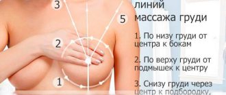
Try to find out if women in your family have had breast problems. If yes, urgently learn self-examination techniques. Periodically feel your breasts to identify the presence of lumps, nodules, roughness on the skin, and soreness. Self-examination should be carried out once a month (and on the same days!). If you find something unclear, consult a doctor immediately. You can perform a breast self-examination: Lying down. Place a pillow under your right shoulder blade and raise your arm above your head. First, examine the right breast (with the fingers of your left hand) in a circular motion with light pressure, starting from the edges of the mammary gland towards the nipple.
In the shower. Raise your right hand. With the fingers of your left hand, feel all parts of the right mammary gland, paying attention to whether there are any swelling or lumps. The left breast is examined in the same way.
At the mirror. Examine the chest, first lowering your hands and then raising them. Pay attention to any enlargements, changes in skin color or shape. Gently squeeze both nipples at the base and make sure there is no colorless or bloody discharge.
Treatment of ulcerative formations on the breast and nipples
Therapy for mammary ulcers depends on the nature of the disease that provoked this phenomenon. Based on the diagnostic results, the following treatment methods are used:
- Medication (conservative).
- Surgical.
The use of therapy is complex, in compliance with the following rules:
- Relief of external phenomena using topical medications.
- Strengthening the immune system with vitamin complexes.
- Elimination of the provoking factor.
Conservative treatment
During drug therapy, manipulations are performed to ensure the outflow of liquid exudate from the ulcerative area of the chest into the bandage.
For this purpose, compresses soaked in sodium chloride solution are applied to the painful area, which:
- Clears the ulcer.
- Improves nutrition of healthy tissues.
- Promotes the formation of granulations.
After the painful area is cleaned and filled with granulations, ointment and gel medications are used, depending on whether the ulcer is wet or dry:
- Eplan.
- Bepanten.
- Solcoseryl.
- Celestoderm.
Solcoseryl ointment has an effective medicinal effect:
- Improves metabolic processes in damaged tissues.
- Accelerates the healing process of the painful area.
In addition, it is recommended:
| For pain relief | For fungal ulcers |
| Levomekol. | Mycozoral, Nizorex, Sebozol. |
Methods of conservative treatment may differ, depending on the origin of the provoking factor.
Tuberculous ulcer of the breast
The use of anti-tuberculosis drugs at an early stage of the disease has a fairly effective effect. For this purpose the following are used:
- Isoniazid.
- Ethambutol.
- Rifampicin.
In severe cases, sectoral resection of the chest area affected by tuberculosis is performed.
Additional methods used:
- UHF.
- Ural Federal District.
- Ultrasound.
- Electrophoresis.
- Inductothermy.
- Magnetotherapy.
Trophic ulcer
The disease requires a comprehensive treatment approach. To eliminate pathology, the following are prescribed:
| Group of drugs | Example of drugs |
| Angioprotectors and disaggregants | Heparin, Prostaglandins, Pentoxifylline, Acetylsalicylic acid. |
| Antibacterial agents | Fuzidin, Levomycetin, Miramistin and Hexicon. |
| Wound healing | Ebermin, Sulfargin and Actovegin. |
In addition to medications, ulcers are washed with sterile saline.
In severe cases, surgery is used to remove necrotic areas, abnormal discharge and damaged tissue. After surgery, ointments with antibacterial or regenerative effects are applied.
To speed up wound healing, physiotherapy is recommended:
- Magnetotherapy.
- UV irradiation.
- Laser therapy.
- Ultrasound cavitation of wounds.
- Hyperbaric oxygenation.
Ulcers under the bust area
Ulcers under the breasts are eliminated with topical medications and other means with healing properties:
- Teymurov ointment.
- Zinc ointment.
- Linin.
- Sea buckthorn oil.
- Creams based on calendula.
- Desyatin (baby cream).
- Tar ointment (for disinfection).
To heal wounds, the following are prescribed:
- Levosin.
- Levomekol.
In severe cases, hormonal medications are indicated:
- Lorinden.
- Solcoseryl.
- Panthenol.
Thoracic herpes
Features of treatment of the sore depend on the type of provoking virus and the stage of the pathological condition. First of all, actions are taken to eliminate the viral irritant from the body. Medicines prescribed:
- Antiviral.
- Anti-inflammatory.
- Corticosteroids.
- Vitamin complexes.
- Antihistamines.
For ulcers of seborrheic or atopic dermatitis, the use of external medications is indicated:
- La Cree.
- Calamine ointment.
- Salicylic-zinc ointment.
- Hydrocortisone cream.
For allergic dermatitis, the following remedies are recommended:
- Anti-allergenic.
- Hormonal.
- Plant based.
Surgical intervention
Surgical treatment is indicated in cases where an ulcer on the mammary gland is a consequence of:
- Purulent mastitis.
- Malignant tumor.
Purulent mastitis
The abscess is opened and the purulent contents are removed. If there is a necrotic process in the breast tissue, a course of antibiotics is prescribed. If a woman is breastfeeding, then breastfeeding is curtailed during treatment.
RMJ
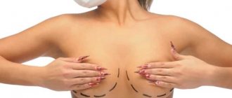
In advanced-stage oncology, specialists try to preserve, as much as possible, as much healthy breast tissue as possible, excising only pathological tumors. However, if it is impossible to preserve it, complete amputation of the diseased breast is performed. After this, mammoplasty is performed.
Drug treatment of ulcerative formations in the cancer process involves the use of:
- Hormone therapy.
- Chemotherapy.
- Radiation therapy.
- Palliative care.
Folk recipes
Experts do not deny the possibility of using alternative medicine recipes for ulcerative formations on the breast, but the advisability of their use depends on the nature of their root cause.
If the ulcers are a consequence of advanced mastitis or the disintegration of a malignant tumor, then only intensive therapy with the use of specific medications and surgery can alleviate the woman’s condition.
It is advisable to use folk remedies for other pathological situations caused by herpes, dermatitis, injuries and only with the permission of a doctor.
| Recipes | Features of application |
| Boron-lemon mixture | Add 6 drops of boric acid and lemon juice to 50 g of cooled boiled water. Soak gauze and apply to damaged areas. |
| Herbal bath | Used for dermatitis. For a therapeutic bath you will need: mint, string, chamomile, oregano - 1 tablespoon each. Combine the ingredients, pour boiling water, leave for at least an hour and strain. Take a bath so that the affected breast is completely in the medicinal water. |
| Black tea with mint | Prepare strong tea with mint and let it brew. Pour into ice cube trays and freeze. Apply the finished cubes to ulcerative lesions. It is used for ulcers under the bust to: - reduce itchy discomfort; - eliminating burning sensation; - temperature decrease. |
| Honey | Shows good antiseptic properties. Apply honey to sore areas for 10-15 minutes, 2 times a day. |
| Lotions from celandine and bran | Steam the bran and celandine. Apply 2 times a day for 15-20 minutes. After the procedure, rinse the breast with warm water. |
| Decoction of chamomile and string | Take 1 tbsp per glass of boiling water. strings and daisies. It is allowed to add mineral water in a 1:1 ratio. Wipe the affected area with the prepared liquid. |
Go to a doctor
If during a self-examination you find a lump or notice discharge from the nipple, contact a mammologist. The accuracy of diagnosis by an experienced specialist is 90-95%. When examining the mammary glands, the doctor usually uses a “diagnostic triangle”: examination, visualization of the condition (mammography and ultrasound) and, if necessary, a biopsy (a tiny piece of tissue is taken and examined under a microscope). If during the examination the mammologist finds discharge from the nipple, he can take a smear and send it to the laboratory for examination. The second stage of the preventive examination is ultrasound and mammography. By the way, the last method is much more effective. Mammography is an x-ray examination that can detect even a small formation that is not detectable upon examination and palpation.
Stages of breast cancer:
- Zero stage. The tumor has not spread to other organs except the breast. This stage gives a ten-year survival rate in almost 100% of cases.
- First stage. The tumor is small, maximum diameter 2 cm, and has not spread to other tissues. with treatment, the ten-year survival rate is about 95–96%.
- Second stage (2A and 2B). Tumor size less than 2 cm, spread to 1–3 lymph nodes, or tumor size 5 cm, but no spread to lymph nodes (2A). Tumor size less than 5 cm, spread to 1–3 lymph nodes, or tumor size more than 5 cm, but without lymph node involvement (2B). Ten-year survival rate, with treatment, is from 70 to 90%.
- Third stage:
- tumor diameter less than 5 cm, damage to 4–9 lymph nodes (3A). Ten-year survival rate is 65 – 75%.
- the tumor reaches the skin or chest. (3B). Survival rate 10-40%
- tumor involvement of the axillary lymph nodes. survival rate is no more than 9% (3C).
- Fourth stage. Spread of metastases to other organs. Survival rate is less than 9%.